Vascular responses disrupted by fructose-induced hyperinsulinemia improved with delta-9- tetrahydrocannabinol

“Objectives: In recent years, cannabinoids have been shown to have beneficial effects on diabetic vascular complications.

Vascular complications due to fructose-induced hyperinsulinemia (HI) and diabetic vascular complications have similar mechanisms.

The aim of this experimental study was to observe whether the cannabinoid agonist delta-9-tetrahydrocannabinol (THC) has an ameliorating effect on fructose-induced HI and vascular responses in the aortic ringof rats with HI.

Methods: A total of 24 rats were categorized into 4 groups: control (standard food pellets and water), HI (water containing 10% fructose provided for 12 weeks), THC (1.5 mg/kg/day intraperitoneal administration for 4 weeks), and THC+HI.Body weight was measured again on the last day of the study and the serum insulin level was measured with an enzyme-linked immunosorbent assay. The acetylcholine (ACh) maximum relaxant effect in aortic rings pre-contractedwith noradrenaline (NA) was evaluated.

Results: The body weight of THC and THC+HI groups was lower compared with that of the controls (p<0.01). Increasedinsulin level as a result of fructose consumption decreased with THC administration (p<0.01) while the glucose level increased in all other groups compared with the control group (p<0.01, p<0.05). The NA Emax value decreased in thegroup receiving THC treatment (p<0.01). The increased ACh pD2 value in the HI groups also decreased in the THCtreatment group (p<0.0001). The decreased maximum inhibition value in the HI group increased significantly with THC administration (p<0.001).

Conclusion: THC demonstrated beneficial effects on fructose-induced HI. THC improved ACh-induced endothelialdependent relaxation in HI rat aortic rings.”

http://acikerisim.demiroglu.bilim.edu.tr:8080/xmlui/handle/11446/4516

https://internationalbiochemistry.com/jvi.aspx?un=IJMB-83703&volume=

The protective effects of Δ 9 -tetrahydrocannabinol against inflammation and oxidative stress in rat liver with fructose-induced hyperinsulinemia

“Objectives: A large amount of fructose is metabolized in the liver and causes hepatic functional damage. Δ9 -tetrahydrocannabinol (THC) is known as a therapeutic agent for clinical and experimental applications. The study aims to investigate the effects of THC treatment on inflammation, lipid profiles and oxidative stress in rat liver with hyperinsulinemia.

Methods: Sprague-Dawley rats were divided into groups: control, fructose (10% fructose in drinking water for 12 weeks), THC (1.5 mg/kg/day for the last 4 weeks, intraperitoneally) and fructose+THC groups. Biochemical parameters were measured spectrophotometrically. ELISA method was used for insulin measurement. Apoptosis and inflammation markers were detected by the streptavidin-biotin peroxidase method.

Key findings: The consumptions of food and fluid are inversely proportional to fructose and non-fructose groups. Insulin levels were the highest in fructose group. The reduced glutathione-S-transferase level significantly increased in fructose + THC group compared with fructose group. Total cholesterol level in the fructose + THC group was higher than the fructose group. Caspase-3 and NF-κβ immunopositive cell numbers increased in fructose + THC rats compared with fructose group. The number of IL-6 immunopositive cell decreased in fructose + THC group compared with fructose group.

Conclusions: According to the result, long-term and low-dose THC administration may reduce hyperinsulinemia and inflammation in rats to some extent.”

https://pubmed.ncbi.nlm.nih.gov/30427077/

“Taken together, the findings from this study demonstrate that fructose consumption induces hyperinsulinemia, oxidative stress and inflammation in rats. The long-term and low-dose THC administration may prevent hyperinsulinemia and inflammation to some extent.”

https://academic.oup.com/jpp/article/71/3/408/6122105?login=false


Receptor-targeted nanoparticles modulate cannabinoid anticancer activity through delayed cell internalization

Scientific Reports

“Δ9-tetrahydrocannabinol (Δ9-THC) is known for its antitumor activity and palliative effects.

However, its unfavorable physicochemical and biopharmaceutical properties, including low bioavailability, psychotropic side effects and resistance mechanisms associated to dosing make mandatory the development of successful drug delivery systems.

In this work, transferring (Tf) surface-modified Δ9-THC-loaded poly(lactide-co-glycolic) nanoparticles (Tf-THC-PLGA NPs) were proposed and evaluated as novel THC-based anticancer therapy. Furthermore, in order to assess the interaction of both the nanocarrier and the loaded drug with cancer cells, a double-fluorescent strategy was applied, including the chemical conjugation of a dye to the nanoparticle polymer along with the encapsulation of either a lipophilic or a hydrophilic dye.

Tf-THC PLGA NPs exerted a cell viability decreased down to 17% vs. 88% of plain nanoparticles, while their internalization was significantly slower than plain nanoparticles. Uptake studies in the presence of inhibitors indicated that the nanoparticles were internalized through cholesterol-associated and clathrin-mediated mechanisms.

Overall, Tf-modification of PLGA NPs showed to be a highly promising approach for Δ9-THC-based antitumor therapies, potentially maximizing the amount of drug released in a sustained manner at the surface of cells bearing cannabinoid receptors.”

https://pubmed.ncbi.nlm.nih.gov/35079042/

“The potential therapeutic applications of marijuana, firstly reported in 1997 by the National Institutes of Health (NIH, USA), are attributed to a great extent to its main component, Δ9-tetrahydrocannabinol (Δ9-THC)1. This cannabinoid continues to attract special attention in oncology due to its palliative effects and antitumor activity; Δ9-THC has been reported to inhibit tumor angiogenesis and cell growth in malignant tissues, leading to cell death.”

“Δ9-THC has been reported to inhibit tumor angiogenesis and cell growth in malignant tissues.”

“Overall, Tf-modification of PLGA NPs seemed a highly promising approach for Δ9-THC-based antitumor therapies, aiming at a prolonged action of the carrier at the target cell surface. Moreover, the translation of this strategy to the delivery of alternative active pharmaceutical ingredients with pharmacological targets on the surface of cells could lead to advances in related therapies.”

https://www.nature.com/articles/s41598-022-05301-z

In Vitro Effect of Δ9-Tetrahydrocannabinol and Cannabidiol on Cancer-Associated Fibroblasts Isolated from Lung Cancer

ijms-logo

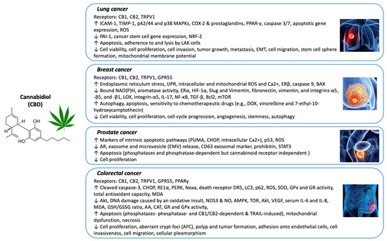
“There is evidence that demonstrates the effect of cannabinoid agonists inhibiting relevant aspects in lung cancer, such as proliferation or epithelial-to-mesenchymal transition (EMT).

Most of these studies are based on evidence observed in in vitro models developed on cancer cell lines. These studies do not consider the complexity of the tumor microenvironment (TME). One of the main components of the TME is cancer-associated fibroblasts (CAFs), cells that are relevant in the control of proliferation and metastasis in lung cancer.

In this work, we evaluated the direct effects of two cannabinoid agonists, tetrahydrocannabinol (THC) and cannabidiol (CBD), used alone or in combination, on CAFs and non-tumor normal fibroblasts (NFs) isolated from adenocarcinoma or from healthy lung tissue from the same patients.

We observed that these compounds decrease cell density in vitro and inhibit the increase in the relative expression of type 1 collagen (COL1A1) and fibroblast-specific protein 1 (FSP1) induced by transforming growth factor beta (TGFβ). On the other hand, we studied whether THC and CBD could modulate the interactions between CAFs or NFs and cancer cells. We conditioned the culture medium with stromal cells treated or not with THC and/or CBD and cultured A549 cells with them.

We found that culture media conditioned with CAFs or NFs increased cell density, induced morphological changes consistent with EMT, inhibited cadherin-1 (CDH1) gene expression, and induced an increase in the relative expression of cadherin-2 (CDH2) and vimentin (VIM) genes in A549 cells. These changes were inhibited or decreased by THC and CBD administered alone or in combination. In another series of experiments, we conditioned culture media with A549 cells treated or not with THC and/or CBD, in the presence or absence of TGFβ. We observed that culture media conditioned with A549 in the presence of TGFβ induced an increase in the expression of COL1A1 and VIM, both in CAFs and in non-tumor NFs. Both THC and CBD ameliorated these effects.

In summary, the results presented here reinforce the usefulness of cannabinoid agonists for the treatment of some relevant aspects of lung cancer pathology, and demonstrate in a novel way their possible effects on CAFs as a result of their relationship with cancer cells. Likewise, the results reinforce the usefulness of the combined use of THC and CBD, which has important advantages in relation to the possibility of using lower doses, thus minimizing the psychoactive effects of THC.”

https://pubmed.ncbi.nlm.nih.gov/35743206/

https://www.mdpi.com/1422-0067/23/12/6766


Cannabidiol Interacts Antagonistically with Cisplatin and Additively with Mitoxantrone in Various Melanoma Cell Lines-An Isobolographic Analysis

ijms-logo

“The medical application of cannabidiol (CBD) has been gathering increasing attention in recent years. This non-psychotropic cannabis-derived compound possesses antiepileptic, antipsychotic, anti-inflammatory and anxiolytic properties. Recent studies report that it also exerts antineoplastic effects in multiple types of cancers, including melanoma.

In this in vitro study we tried to reveal the anticancer properties of CBD in malignant melanoma cell lines (SK-MEL 28, A375, FM55P and FM55M2) administered alone, as well as in combination with mitoxantrone (MTX) or cisplatin (CDDP).

The effects of CBD on the viability of melanoma cells were measured by the MTT assay; cytotoxicity was determined in the LDH test and proliferation in the BrdU test. Moreover, the safety of CBD was tested in human keratinocytes (HaCaT) in LDH and MTT tests.

Results indicate that CBD reduces the viability and proliferation of melanoma-malignant cells and exerts additive interactions with MTX. Unfortunately, CBD produced antagonistic interaction when combined with CDDP. CBD does not cause significant cytotoxicity in HaCaT cell line.

In conclusion, CBD may be considered as a part of melanoma multi-drug therapy when combined with MTX. A special attention should be paid to the combination of CBD with CDDP due to the antagonistic interaction observed in the studied malignant melanoma cell lines.”

https://pubmed.ncbi.nlm.nih.gov/35743195/

https://www.mdpi.com/1422-0067/23/12/6752

Mechanisms of Cannabidiol (CBD) in Cancer Treatment: A Review

biology-logo


“Cannabis sativa L. (Cannabis) and its bioactive compounds, including cannabinoids and non-cannabinoids, have been extensively studied for their biological effects in recent decades. Cannabidiol (CBD), a major non-intoxicating cannabinoid in Cannabis, has emerged as a promising intervention for cancer research.

The purpose of this review is to provide insights into the relationship between CBD and cancer based on recent research findings.

The anticancer effects of CBD are mainly mediated via its interaction with the endocannabinoid system, resulting in the alleviation of pain and the promotion of immune regulation. Published reviews have focused on the applications of CBD in cancer pain management and the possible toxicological effects of its excessive consumption.

In this review, we aim to summarize the mechanisms of action underlying the anticancer activities of CBD against several common cancers. Studies on the efficacy and mechanisms of CBD on cancer prevention and intervention in experimental models (i.e., cell culture- and animal-based assays) and human clinical studies are included in this review.”

https://pubmed.ncbi.nlm.nih.gov/35741337/

“Emerging evidence suggests positive outcomes from the use of CBD as a cancer treatment. CBD can relieve cancer pain and ease the side effects of chemotherapy; however, there is less research about the mechanism of CBD’s anticancer effects. In this article, recent studies on the efficacy and mechanisms of CBD’s anticancer effects in cell- and animal-based models and human clinical studies are reviewed.”

https://www.mdpi.com/2079-7737/11/6/817


Cannabidiol and Cannabigerol Inhibit Cholangiocarcinoma Growth In Vitro via Divergent Cell Death Pathways

biomolecules-logo

“Cholangiocarcinoma (CCA) is a rare and highly lethal disease with few effective treatment options.

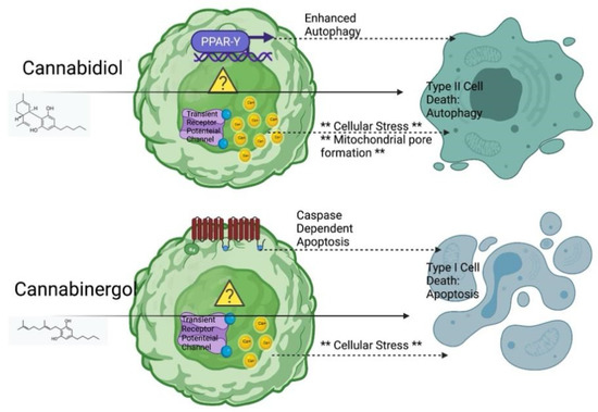
Cannabinoids, cannabidiol (CBD) and cannabigerol (CBG) are non-psychedelic components extracted from cannabis. These non-psychoactive compounds have shown anti-proliferative potential in other tumor models; however, the efficacy of CBD and CBG in CCA is unknown. Furthermore, two cell death pathways are implicated with CBD resulting in autophagic degeneration and CBG in apoptosis. HuCC-T1 cells, Mz-ChA-1 cells (CCA cell lines) and H69 cells (immortalized cholangiocytes), were treated with CBD and CBG for 24 to 48 h.

The influence of these cannabinoids on proliferation was assessed via MTT assay. Apoptosis and cell cycle were evaluated via Annexin-V apoptosis assay and propidium iodide, respectively. The expression of proliferation biomarker Ki-67, apoptosis biomarker BAX, and autophagic flux biomarkers LC3b and LAMP1 were evaluated via immunofluorescence. Cell migration and invasion were evaluated via wound healing assay and trans-well migration invasion assays, respectively. The colony formation was evaluated via colony formation assay. In addition, the expression of autophagy gene LC3b and apoptosis genes BAX, Bcl-2, and cleaved caspase-3 were evaluated via Western blot.

CBD and CBG are non-selective anti-proliferative agents yielding similar growth curves in CCA; both cannabinoids are effective, yet CBG is more active at lower doses. Low doses of CBD and CBG enhanced immortalized cholangiocyte activity. The reduction in proliferation begins immediately and occurs maximally within 24 h of treatment. Moreover, a significant increase in the late-stage apoptosis and a reduction in the number of cells in S stage of the cell cycle indicates both CBD and CBG treatment could promote apoptosis and inhibit mitosis in CCA cells. The fluorescent expression of BAX and LC3b was significantly enhanced with CBD treatment when compared to control. LAMP1 and LC3b colocalization could also be observed with CBD and CBG treatment indicating changes in autophagic flux.

A significant inhibition of migration, invasion and colony formation ability was shown in both CBD and CBG treatment in CCA. Western blot showed an overall decrease in the ratio of anti-apoptotic protein Bcl-2 with respect to pro-apoptotic protein BAX with CBG treatment. Furthermore, CBD treatment enhanced the expression of Type II cell death (autophagic degeneration) protein LC3b, which was reduced in CBG-treated CCA cells. Meanwhile, CBG treatment upregulated Type I cell death (programmed apoptosis) protein cleaved caspase-3.

CBD and CBG are effective anti-cancer agents against CCA, capable of inhibiting the classic hallmarks of cancer, with a divergent mechanism of action (Type II or Type I respectively) in inducing these effects.”

https://pubmed.ncbi.nlm.nih.gov/35740979/

https://www.mdpi.com/2218-273X/12/6/854


Biosynthetic origins of unusual cannabimimetic phytocannabinoids in Cannabis sativa L: A review

Phytochemistry

“Plants of Cannabis sativa L. (Cannabaceae) produce an array of more than 160 isoprenylated resorcinyl polyketides, commonly referred to as phytocannabinoids. These compounds represent molecules of therapeutic importance due to their modulation of the human endocannabinoid system (ECS).

While understanding of the biosynthesis of the major phytocannabinoids Δ9-tetrahydrocannabinol (Δ9-THC) and cannabidiol (CBD) has grown rapidly in recent years, the biosynthetic origin and genetic regulation of many potentially therapeutically relevant minor phytocannabinoids remains unknown, which limits the development of chemotypically elite varieties of C. sativa.

This review provides an up-to-date inventory of unusual phytocannabinoids which exhibit cannabimimetic-like activities and proposes putative metabolic origins. Metabolic branch points exploitable for combinatorial biosynthesis and engineering of phytocannabinoids with augmented therapeutic activities are also described, as is the role of phytocannabinoid remodelling to accelerate therapeutic portfolio expansion in C. sativa.”

https://pubmed.ncbi.nlm.nih.gov/35718133/

https://www.sciencedirect.com/science/article/abs/pii/S0031942222001984?via%3Dihub

Phytocannabinoids and Cannabis-Based Products as Alternative Pharmacotherapy in Neurodegenerative Diseases: From Hypothesis to Clinical Practice

Archive of "Frontiers in Cellular Neuroscience". - PMC

“Historically, Cannabis is one of the first plants to be domesticated and used in medicine, though only in the last years the amount of Cannabis-based products or medicines has increased worldwide.

Previous preclinical studies and few published clinical trials have demonstrated the efficacy and safety of Cannabis-based medicines in humans. Indeed, Cannabis-related medicines are used to treat multiple pathological conditions, including neurodegenerative disorders.

In clinical practice, Cannabis products have already been introduced to treatment regimens of Alzheimer’s disease, Parkinson’s disease and Multiple Sclerosis’s patients, and the mechanisms of action behind the reported improvement in the clinical outcome and disease progression are associated with their anti-inflammatory, immunosuppressive, antioxidant, and neuroprotective properties, due to the modulation of the endocannabinoid system.

In this review, we describe the role played by the endocannabinoid system in the physiopathology of Alzheimer, Parkinson, and Multiple Sclerosis, mainly at the neuroimmunological level. We also discuss the evidence for the correlation between phytocannabinoids and their therapeutic effects in these disorders, thus describing the main clinical studies carried out so far on the therapeutic performance of Cannabis-based medicines.”

https://pubmed.ncbi.nlm.nih.gov/35707521/

“Based on scientific evidence, the use of Cannabis-based products or Cannabis-based medicine (CBM) has been growing among patients diagnosed with neurodegenerative diseases. Most reports of clinical trials also describe significant improvement in disease-related primary and/or secondary symptoms, besides general improvement in life quality.”

https://www.frontiersin.org/articles/10.3389/fncel.2022.917164/full


Cannabinoids as Glial Cell Modulators in Ischemic Stroke: Implications for Neuroprotection

Frontiers in Pharmacology welcomes new Field Chief Editor – Science &  research news | Frontiers

“Stroke is the second leading cause of death worldwide following coronary heart disease. Despite significant efforts to find effective treatments to reduce neurological damage, many patients suffer from sequelae that impair their quality of life. For this reason, the search for new therapeutic options for the treatment of these patients is a priority.

Glial cells, including microglia, astrocytes and oligodendrocytes, participate in crucial processes that allow the correct functioning of the neural tissue, being actively involved in the pathophysiological mechanisms of ischemic stroke. Although the exact mechanisms by which glial cells contribute in the pathophysiological context of stroke are not yet completely understood, they have emerged as potentially therapeutic targets to improve brain recovery.

The endocannabinoid system has interesting immunomodulatory and protective effects in glial cells, and the pharmacological modulation of this signaling pathway has revealed potential neuroprotective effects in different neurological diseases. Therefore, here we recapitulate current findings on the potential promising contribution of the endocannabinoid system pharmacological manipulation in glial cells for the treatment of ischemic stroke.”

https://pubmed.ncbi.nlm.nih.gov/35721207/

“In summary, due to the profound implication of glial cells in stroke, the pharmacological modulation of the glial endocannabinoid system (ECS) could represent a significant advantage to help reduce/limit neuronal damage and stroke-associated sequelae.”

https://www.frontiersin.org/articles/10.3389/fphar.2022.888222/full