Targeting dysfunctional endocannabinoid signaling in a mouse model of Gulf War illness

pubmed logo

“Gulf War Illness (GWI) is a chronic disorder characterized by a heterogeneous set of symptoms that include pain, fatigue, anxiety, and cognitive impairment. These are thought to stem from damage caused by exposure under unpredictable stress to toxic Gulf War (GW) chemicals, which include pesticides, nerve agents, and prophylactic drugs.

We hypothesized that GWI pathogenesis might be rooted in long-lasting disruption of the endocannabinoid (ECB) system, a signaling complex that serves important protective functions in the brain.

Using a mouse model of GWI, we found that tissue levels of the ECB messenger, anandamide, were significantly reduced in the brain of diseased mice, compared to healthy controls. In addition, transcription of the Faah gene, which encodes for fatty acid amide hydrolase (FAAH), the enzyme that deactivates anandamide, was significant elevated in prefrontal cortex of GWI mice and brain microglia.

Behavioral deficits exhibited by these animals, including heightened anxiety-like and depression-like behaviors, and defective extinction of fearful memories, were corrected by administration of the FAAH inhibitor, URB597, which normalized brain anandamide levels. Furthermore, GWI mice displayed unexpected changes in the microglial transcriptome, implying persistent dampening of homeostatic surveillance genes and abnormal expression of pro-inflammatory genes upon immune stimulation.

Together, these results suggest that exposure to GW chemicals produce a deficit in brain ECB signaling which is associated with persistent alterations in microglial function. Pharmacological normalization of anandamide-mediated ECB signaling may offer an effective therapeutic strategy for ameliorating GWI symptomology.”

https://pubmed.ncbi.nlm.nih.gov/39241906/

“A mouse model for Gulf War Illness (GWI) displays deficits in brain anandamide.

Normalization of endocannabinoid signaling may offer a therapeutic strategy for GWI.”

https://www.sciencedirect.com/science/article/pii/S0028390824003113?via%3Dihub

“FDA-approved cannabidiol [Epidiolex®] alleviates Gulf War Illness-linked cognitive and mood dysfunction, hyperalgesia, neuroinflammatory signaling, and declined neurogenesis”

https://pubmed.ncbi.nlm.nih.gov/39169440/

“CBD formulation improves energetic homeostasis in dermal fibroblasts from Gulf War Illness patients.  Our data provide new evidence that will validate the potential of cannabinoids as a therapeutic strategy to mitigate energy imbalance that may contribute to detrimental symptomatology (i.e., chronic fatigue, brain fog, cognitive dysfunction, etc.) in GWI patients.”

https://pubmed.ncbi.nlm.nih.gov/35560565/

The Endocannabinoid System in Alzheimer’s Disease: A Network Meta-Analysis

pubmed logo

“The findings concerning the association between endocannabinoid system (ECS) and Alzheimer’s disease (AD) exhibited inconsistencies when examining the expression levels of endocannabinoids. This study aimed to provide a comprehensive summary of the studies regarding alterations of the ECS in AD.

Six databases were thoroughly searched for literature to select relevant studies investigating the ECS in AD, including changes in cannabinoid receptors (CB1R and CB2R), endocannabinoids (2-AG and AEA), and their associated enzymes (FAAH and MAGL). Traditional meta-analysis evaluated the expression levels of the ECS in AD, and the results showed no significant differences in ECS components between healthy controls and AD patients. However, subgroup analysis revealed significantly lower expression levels of CB1R in AD than in controls, particularly in studies using western blot (SMD = -0.88, p < 0.01) and in studies testing CB1R of frontal cortex (SMD = -1.09, p < 0.01). For studies using HPLC, the subgroup analysis indicated significantly higher 2-AG levels in AD than in controls (SMD = 0.46, p = 0.02). Network meta-analysis examined the rank of ECS alterations in AD compared to controls, and the findings revealed that 2-AG and MAGL exhibited the largest increase and CB1R showed the largest decrease relative to the control group.

Based on the findings of traditional meta-analysis and network meta-analysis, we proposed that AD patients may present decreased expression levels of CB1R and increased expression levels of 2-AG and its degrading enzyme MAGL.

Our results may contribute to the growing body of research supporting the therapeutic potential of ECS modulation in the management of AD.”

https://pubmed.ncbi.nlm.nih.gov/39245959/

https://onlinelibrary.wiley.com/doi/10.1002/jnr.25380

Microglial morphological/inflammatory phenotypes and endocannabinoid signaling in a preclinical model of periodontitis and depression

pubmed logo

“Background: Depression is a chronic psychiatric disease of multifactorial etiology, and its pathophysiology is not fully understood. Stress and other chronic inflammatory pathologies are shared risk factors for psychiatric diseases, and comorbidities are features of major depression. Epidemiological evidence suggests that periodontitis, as a source of low-grade chronic systemic inflammation, may be associated with depression, but the underlying mechanisms are not well understood.

Methods: Periodontitis (P) was induced in Wistar: Han rats through oral gavage with the pathogenic bacteria Porphyromonas gingivalis and Fusobacterium nucleatum for 12 weeks, followed by 3 weeks of chronic mild stress (CMS) to induce depressive-like behavior. The following four groups were established (n = 12 rats/group): periodontitis and CMS (P + CMS+), periodontitis without CMS, CMS without periodontitis, and control. The morphology and inflammatory phenotype of microglia in the frontal cortex (FC) were studied using immunofluorescence and bioinformatics tools. The endocannabinoid (EC) signaling and proteins related to synaptic plasticity were analyzed in FC samples using biochemical and immunohistochemical techniques.

Results: Ultrastructural and fractal analyses of FC revealed a significant increase in the complexity and heterogeneity of Iba1 + parenchymal microglia in the combined experimental model (P + CMS+) and increased expression of the proinflammatory marker inducible nitric oxide synthase (iNOS), while there were no changes in the expression of cannabinoid receptor 2 (CB2). In the FC protein extracts of the P + CMS + animals, there was a decrease in the levels of the EC metabolic enzymes N-acyl phosphatidylethanolamine-specific phospholipase D (NAPE-PLD), diacylglycerol lipase (DAGL), and monoacylglycerol lipase (MAGL) compared to those in the controls, which extended to protein expression in neurons and in FC extracts of cannabinoid receptor 1 (CB1) and to the intracellular signaling molecules phosphatidylinositol-3-kinase (PI3K), protein kinase B (Akt) and extracellular signal-regulated kinase 1/2 (ERK1/2). The protein levels of brain-derived neurotrophic factor (BDNF) and synaptophysin were also lower in P + CMS + animals than in controls.

Conclusions: The combined effects on microglial morphology and inflammatory phenotype, the EC signaling, and proteins related to synaptic plasticity in P + CMS + animals may represent relevant mechanisms explaining the association between periodontitis and depression. These findings highlight potential therapeutic targets that warrant further investigation.”

https://pubmed.ncbi.nlm.nih.gov/39245706/

https://jneuroinflammation.biomedcentral.com/articles/10.1186/s12974-024-03213-5

Cannabidiol suppresses silica-induced pulmonary inflammation and fibrosis through regulating NLRP3/TGF-β1/Smad2/3 pathway

pubmed logo

“Silica-induced pulmonary fibrosis is an irreversible and progressive lung disease with limited treatments available. In this work, FDA-approved cannabidiol (CBD) was studied for its potential medical use in silicosis.

In silicosis female C57BL/6 mice model, oral CBD or pirfenidone (PFD) on day 1 after intratracheal drip silica (150 mg/mL) and continued for 42 days. Lung inflammatory and fibrotic changes were studied using ELISA kits, H&E staining and Masson staining. Osteopontion (OPN) and α-smooth muscle actin (α-SMA) expression in lung tissues was determined using immunohistochemical staining.

The results indicated that CBD attenuated silica-induced pulmonary inflammation and fibrosis.

Human myeloid leukemia mononuclear cells (THP-1) were treated with silica (200 μg/mL) to induce cell damage, then CBD (10 μM, 20 μM) and PFD (100 μM) were incubated. In vitro experiments showed that CBD can effectively reduce the expression of NLRP3 inflammasome in THP-1 cells and subsequently block silica-stimulated transformation of fibromuscular-myofibroblast transition (FMT) by culturing human embryonic lung fibroblasts (MRC-5) in conditioned medium of THP-1 cells.

Therefore, CBD exhibited the potential therapy for silicosis through inhibiting the silica-induced pulmonary inflammation and fibrosis via the NLRP3/TGF-β1/Smad2/3 signaling pathway.”

https://pubmed.ncbi.nlm.nih.gov/39244899/

“CBD ameliorates silica-induced pulmonary inflammation and fibrosis.”

https://www.sciencedirect.com/science/article/abs/pii/S1567576924016096?via%3Dihub

“Silica compounds are found throughout the environment in rocks, sand, clay, soil, air, and water. Silica is used in many commercial products, such as bricks, glass and ceramics, plaster, granite, concrete, cleansers, skin care products, and talcum powder. Some forms of amorphous silica are used as food additives, food wrappings, toothpaste and cosmetics. The general population is exposed to silica through air, certain types of indoor dust (such as from concrete), food, water, soil, and some consumer products. The exposure of greatest concern is through air.”

https://wwwn.cdc.gov/TSP/ToxFAQs/ToxFAQsDetails.aspx?faqid=1492&toxid=290#:~:text=Silica%20is%20used%20in%20many,food%20wrappings%2C%20toothpaste%20and%20cosmetics.

Neuromolecular and behavioral effects of Cannabidiol on depressive-associated behaviors and neuropathic pain conditions in mice

pubmed logo

“Background and aims: Neuropathic pain (NP) has a high incidence in the general population, is closely related to anxiety disorders, and has a negative impact on the quality of life. Cannabidiol (CBD), as a natural product, has been extensively studied for its potential therapeutic effects on symptoms such as pain and depression (DP). However, the mechanism of CBD in improving NP with depression is not fully understood.

Methods: First, we used bioinformatics tools to deeply mine the intersection genes associated with NP, DP, and CBD. Secondly, the core targets were screened by Protein-protein interaction network, Gene Ontology, Kyoto Encyclopedia of Genes and Genomes analysis, molecular docking and molecular dynamics simulation. Next, the effects of CBD intervention on pain and depressive behaviors in the spinal nerve ligation (SNL) mouse model were evaluated using behavioral tests, and dose-response curves were plotted. After the optimal intervention dose was determined, the core targets were verified by Western blot (WB) and Quantitative Polymerase Chain Reaction (qPCR). Finally, we investigated the potential mechanism of CBD by Nissl staining, Immunofluorescence (IF) and Transmission Electron Microscopy (TEM).

Results: A total of five core genes of CBD most associated with NP and DP were screened by bioinformatics analysis, including PTGS2, GPR55, SOD1, CYP1A2 and NQO1. Behavioral test results showed that CBD by intraperitoneal administration 5mg/kg can significantly improve the pain behavior and depressive state of SNL mice. WB, qPCR, IF, and TEM experiments further confirmed the regulatory effects of CBD on key molecules.

Conclusion: In this study, we found five targets of CBD in the treatment of NP with DP. These findings provide further theoretical and experimental basis for CBD as a potential therapeutic agent.”

https://pubmed.ncbi.nlm.nih.gov/39245142/

“We identified five core genes associated with comorbid NP and DP targeted by CBD. CBD intervention can improve NP and depressive-associated behavior in mice.”

https://www.sciencedirect.com/science/article/abs/pii/S0028390824003228?via%3Dihub


Effects of Cannabidiol on Bone Quality in Ovariectomized Rats

pubmed logo

“The incidence of osteoporosis and related fractures increases significantly with age, impacting public health and associated costs. Postmenopausal osteoporosis results from increased bone resorption due to decreased estrogen levels.

The endocannabinoid system, especially cannabidiol (CBD), has shown therapeutic potential in modulating bone formation. This study investigated the effects of administration of CBD in rats after the onset of with ovariectomy-induced osteopenia (OVX).

Forty-eight female Sprague‒Dawley rats were divided into four groups (n = 12): OVX + CBD, SHAM + CBD, OVX + vehicle, and SHAM + vehicle. CBD was administered intraperitoneally for 3 weeks. After euthanasia, the bone quality, mechanical properties, and bone microarchitecture of the femurs and lumbar vertebrae were assessed by microcomputed tomography (micro-CT), bone densitometry, mechanical tests, and histological and immunohistochemical analyses.

CBD treatment improved the bone mineral density (BMD) of the lumbar vertebrae and increased the BV/TV% and Tb.N in the femoral neck. There were also improvements in the mechanical properties, such as the maximum force and stiffness of the femurs and vertebrae. CBD significantly increased the bone matrix in osteopenic femurs and vertebrae, Although did not significantly influence the expression of RANKL and OPG, in ovariectomized animals, there was an increase in osteoblasts and a decrease in osteoclasts.

Determining the optimal timing for CBD use in relation to postovariectomy bone loss remains a crucial issue. Understanding when and how CBD can be most effective in preventing or treating bone loss is essential to emphasize the importance of early diagnosis and treatment of osteoporosis. However, further studies are needed to explore in more detail the efficacy and safety of CBD in the treatment of postmenopausal osteoporosis.”

https://pubmed.ncbi.nlm.nih.gov/39245783/

https://link.springer.com/article/10.1007/s00223-024-01281-6

Cannabinoids elicit antidepressant-like behavior and activate serotonergic neurons through the medial prefrontal cortex

pubmed logo

“Preclinical and clinical studies show that cannabis modulates mood and possesses antidepressant-like properties, mediated by the agonistic activity of cannabinoids on central CB1 receptors (CB1Rs). The action of CB1R agonists on the serotonin (5-HT) system, the major transmitter system involved in mood control and implicated in the mechanism of action of antidepressants, remains however poorly understood.

In this study, we demonstrated that, at low doses, the CB1R agonist WIN55,212-2 [R(+)-[2,3-dihydro-5-methyl-3-[(morpholinyl)]pyrrolo[1,2,3-de]-1,4-benzoxazinyl]-(1-naphthalenyl) methanone mesylate] exerts potent antidepressant-like properties in the rat forced-swim test (FST).

This effect is CB1R dependent because it was blocked by the CB1R antagonist rimonabant and is 5-HT mediated because it was abolished by pretreatment with the 5-HT-depleting agent parachlorophenylalanine. Then, using in vivo electrophysiology, we showed that low doses of WIN55,212-2 dose dependently enhanced dorsal raphe nucleus 5-HT neuronal activity through a CB1R-dependent mechanism.

Conversely, high doses of WIN55,212-2 were ineffective in the FST and decreased 5-HT neuronal activity through a CB1R-independent mechanism. The CB1R agonist-induced enhancement of 5-HT neuronal activity was abolished by total or medial prefrontocortical, but not by lateral prefrontocortical, transection. Furthermore, 5-HT neuronal activity was enhanced by the local microinjection of WIN55,212-2 into the ventromedial prefrontal cortex (mPFCv) but not by the local microinjection of WIN55,212-2 into the lateral prefrontal cortex. Similarly, the microinjection of WIN55,212-2 into the mPFCv produced a CB1R-dependent antidepressant-like effect in the FST.

These results demonstrate that CB1R agonists possess antidepressant-like properties and modulate 5-HT neuronal activity via the mPFCv.”

“These results establish that low doses of a CB1R agonist elicit potent antidepressant-like behavior and enhance 5-HT neurotransmission, mediated by CB1R activation in the mPFCv. Conversely, high doses nullify antidepressant-like behavior and markedly attenuate 5-HT neurotransmission, an effect that appears to be instigated by a non-CB1R mechanism.”

https://pubmed.ncbi.nlm.nih.gov/17959812/

Multi-level therapeutic actions of cannabidiol in ketamine-induced schizophrenia psychopathology in male rats

pubmed logo

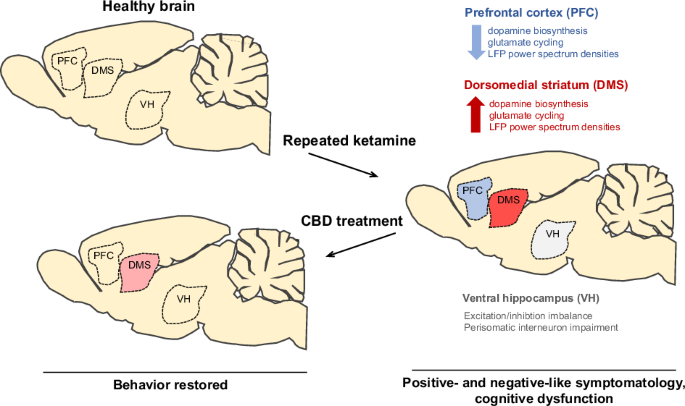
“Repeated administration of ketamine (KET) has been used to model schizophrenia-like symptomatology in rodents, but the psychotomimetic neurobiological and neuroanatomical underpinnings remain elusive. In parallel, the unmet need for a better treatment of schizophrenia requires the development of novel therapeutic strategies.

Cannabidiol (CBD), a major non-addictive phytocannabinoid has been linked to antipsychotic effects with unclear mechanistic basis. Therefore, this study aims to clarify the neurobiological substrate of repeated KET administration model and to evaluate CBD’s antipsychotic potential and neurobiological basis.

CBD-treated male rats with and without prior repeated KET administration underwent behavioral analyses, followed by multilevel analysis of different brain areas including dopaminergic and glutamatergic activity, synaptic signaling, as well as electrophysiological recordings for the assessment of corticohippocampal and corticostriatal network activity.

Repeated KET model is characterized by schizophrenia-like symptomatology and alterations in glutamatergic and dopaminergic activity mainly in the PFC and the dorsomedial striatum (DMS), through a bi-directional pattern. These observations are accompanied by glutamatergic/GABAergic deviations paralleled to impaired function of parvalbumin- and cholecystokinin-positive interneurons, indicative of excitation/inhibition (E/I) imbalance.

Moreover, CBD counteracted the schizophrenia-like behavioral phenotype as well as reverted prefrontal abnormalities and ventral hippocampal E/I deficits, while partially modulated dorsostriatal dysregulations.

This study adds novel insights to our understanding of the KET-induced schizophrenia-related brain pathology, as well as the CBD antipsychotic action through a region-specific set of modulations in the corticohippocampal and costicostrtiatal circuitry of KET-induced profile contributing to the development of novel therapeutic strategies focused on the ECS and E/I imbalance restoration.”

https://pubmed.ncbi.nlm.nih.gov/39242923/

https://www.nature.com/articles/s41386-024-01977-1


Medicinal cannabis extracts are neuroprotective against Aβ1-42-mediated toxicity in vitro

pubmed logo

“Background: Phytocannabinoids inhibit the aggregation and neurotoxicity of the neurotoxic Alzheimer’s disease protein β amyloid (Aβ). We characterised the capacity of five proprietary medical cannabis extracts, heated and non-heated, with varying ratios of cannabidiol and Δ9-tetrahydrocannabinol and their parent carboxylated compounds to protect against lipid peroxidation and Aβ-evoked neurotoxicity in PC12 cells.

Methods: Neuroprotection against lipid peroxidation and Aβ1-42-induced cytotoxicity was assessed using the thiazolyl blue tetrazolium bromide (MTT) assay. Transmission electron microscopy was used to visualise phytocannabinoid effects on Aβ1-42 aggregation and fluorescence microscopy.

Results: Tetrahydrocannabinol (THC)/tetrahydrocannabinolic acid (THCA)-predominant cannabis extracts demonstrated the most significant overall neuroprotection against Aβ1-42-induced loss of PC12 cell viability. These protective effects were still significant after heating of extracts, while none of the extracts provided significant neuroprotection to lipid peroxidation via tbhp exposure. Modest inhibition of Aβ1-42 aggregation was demonstrated only with the non-heated BC-401 cannabis extract, but overall, there was no clear correlation between effects on fibrils and conferral of neuroprotection.

Conclusions: These findings highlight the variable neuroprotective activity of cannabis extracts containing major phytocannabinoids THC/THCA and cannabidiol (CBD)/cannabidiolic acid (CBDA) on Aβ-evoked neurotoxicity and inhibition of amyloid β aggregation. This may inform the future use of medicinal cannabis formulations in the treatment of Alzheimer’s disease and dementia.”

https://pubmed.ncbi.nlm.nih.gov/39243211/

“With access to approved pathways increasing globally, medicinal cannabis formulations are increasingly being used to treat neuropsychiatric conditions. With laboratory and animal studies now showing benefits of cannabis and cannabinoids in treating neurodegenerative diseases, this study investigated whether whole cannabis extracts could protection neuronal cells against the toxicity of a signature Alzheimer’s disease protein, beta (β) amyloid.

We found that cannabis extracts afforded neuronal cells protection against amyloid β toxicity, mostly in extracts with the major phytocannabinoid, Δ9-THC, or its parent compound, Δ9-THC-COOH. These results suggest that medicinal cannabis may have potential in the further treatment of dementia.”

https://onlinelibrary.wiley.com/doi/10.1111/bcpt.14078


A combination of Δ9-tetrahydrocannabinol and cannabidiol modulates glutamate dynamics in the hippocampus of an animal model of Alzheimer’s disease

pubmed logo

“A combination of Δ9-tetrahydrocannabinol (Δ9-THC) and cannabidiol (CBD) at non-psychoactive doses was previously demonstrated to reduce cognitive decline in APP/PS1 mice, an animal model of Alzheimer’s disease (AD). However, the neurobiological substrates underlying these therapeutic properties of Δ9-THC and CBD are not fully understood.

Considering that dysregulation of glutamatergic activity contributes to cognitive impairment in AD, the present study evaluates the hypothesis that the combination of these two natural cannabinoids might reverse the alterations in glutamate dynamics within the hippocampus of this animal model of AD.

Interestingly, our findings reveal that chronic treatment with Δ9-THC and CBD, but not with any of them alone, reduces extracellular glutamate levels and the basal excitability of the hippocampus in APP/PS1 mice.

These effects are not related to significant changes in the function and structure of glutamate synapses, as no relevant changes in synaptic plasticity, glutamate signaling or in the levels of key components of these synapses were observed in cannabinoid-treated mice. Our data instead indicate that these cannabinoid effects are associated with the control of glutamate uptake and/or to the regulation of the hippocampal network.

Taken together, these results support the potential therapeutic properties of combining these natural cannabinoids against the excitotoxicity that occurs in AD brains.”

https://pubmed.ncbi.nlm.nih.gov/39232876/

https://www.neurotherapeuticsjournal.org/article/S1878-7479(24)00126-0/fulltext