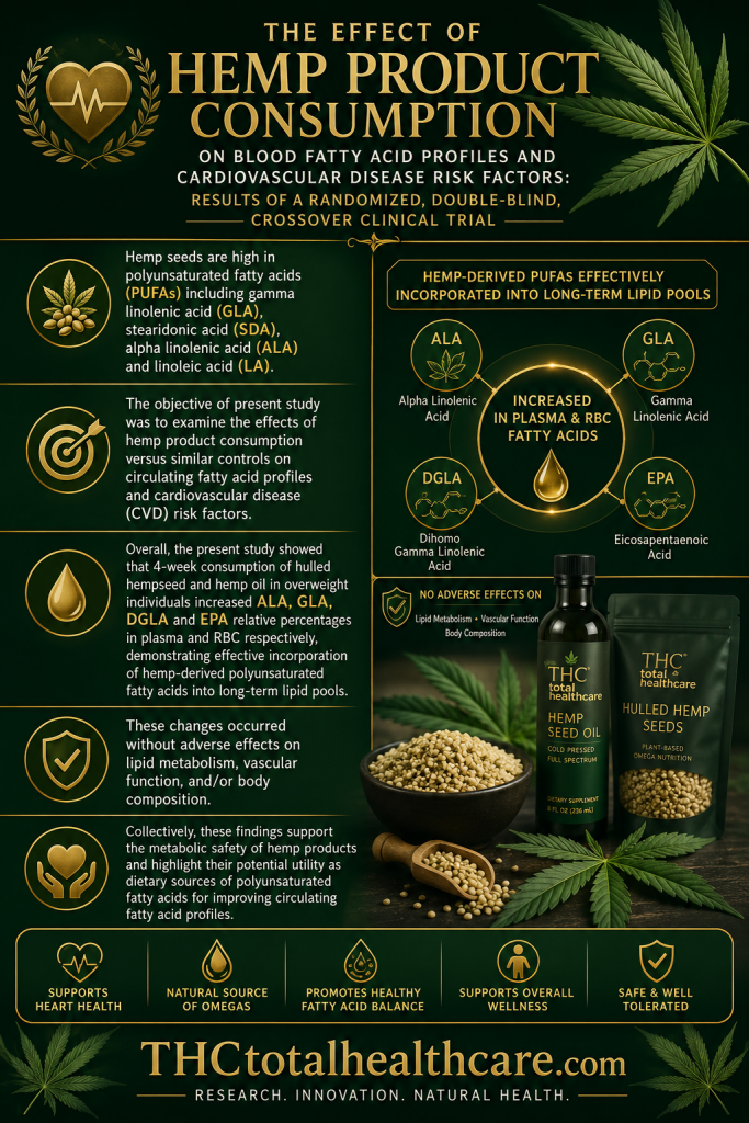
Therapeutic Effect of Lebanese Cannabis Oil Extract in the Management of Sodium Orthovanadate-Induced Nephrotoxicity in Rats

pubmed logo

“Sodium orthovanadate is a non-selective protein tyrosine phosphatase inhibitor that can cause several types of kidney injury, including glomerulosclerosis, inflammation, and tubular damage.

Cannabis is widely known for its medicinal use, and several studies have demonstrated its anti-diabetic and anti-inflammatory properties.

The current study investigated the therapeutic effect of Lebanese cannabis oil extract (COE) against sodium orthovanadate-induced nephrotoxicity both in vitro and in vivo.

Sprague Dawley male rats were intraperitoneally injected with 10 mg/kg sodium orthovanadate for 10 days followed by 5 mg/kg; 10 mg/kg; or 20 mg/kg intraperitoneal injection of cannabis oil extract, starting on day 4 until day 10. The body weight of the rats was monitored during the study, and clinical parameters, including serum urea, creatinine, and electrolytes, as well as kidney and heart pathology, were measured. Conditionally immortalized cultured rat podocytes were exposed to either sodium orthovanadate or selective phosphatase inhibitors, including DUSPi (DUSP1/6 inhibitor) and SF1670 (PTEN inhibitor), in the presence or absence of cannabis oil extract. MTS and an in vitro scratch assay were used to assess podocyte cell viability and migration, respectively. Western blot analysis was used to evaluate the phosphorylation levels of AKT and p38 MAPK.

Rats injected with sodium orthovanadate displayed a marked reduction in body weight and an increase in serum creatinine and urea in comparison to the control non-treated group. All doses of COE caused a significant decrease in serum urea, with a significant decrease in serum creatinine observed at a dose of 20 mg/kg. Moreover, the COE treatment of rats injected with orthovanadate (20 mg/kg) showed a marked reduction in renal vascular dilatation, scattered foci of acute tubular necrosis, and numerous mitoses in tubular cells compared to the sodium orthovanadate-treated group.

The cell viability assay revealed that COE reversed cytotoxicity induced by sodium orthovanadate and specific phosphatase inhibitors (DUSPi and SF1670) in rat podocytes. The in vitro scratch assay showed that COE partially restored the migratory capacity of podocytes incubated with DUSPi and SF1670. Time-course and dose-dependent experiments showed that COE (1 μg/mL) induced a significant increase in phospho-(S473)-AKT, along with a decrease in phospho (T180 + Y182) P38 levels.

The current results demonstrated that Lebanese cannabis oil possesses important kidney protective effects against sodium orthovanadate-induced renal injury.”

https://pubmed.ncbi.nlm.nih.gov/40362381/

https://www.mdpi.com/1422-0067/26/9/4142

Targeting the Gut-Brain Axis with Plant-Derived Essential Oils: Phytocannabinoids and Beyond

pubmed logo

“Background: The gut-brain axis (GBA) is a complex bidirectional communication system that links the gastrointestinal tract and the central nervous system. Essential oils (EOs) have emerged as promising natural compounds capable of modulating this axis. 

Methods: A comprehensive analysis of the recent literature was conducted, focusing on studies investigating the effects of EOs on the GBA.

Particular attention was given to the endocannabinoid system, the role of cannabis-derived EOs, and other plant-based EOs with potential neuroprotective and gut microbiota-modulating effects. 

Results: Among the EOs analyzed, cannabis essential oil (CEO) gained attention for its interaction with cannabinoid receptors (CBR1 and CBR2), modulating gut motility, immune responses, and neurotransmission. While acute administration of the CEO reduces inflammation and gut permeability, chronic use has been associated with alterations in gut microbiota composition, potentially impairing cognitive function. Other EOs, such as those from rosemary, lavender, eucalyptus, and oregano, demonstrated effects on neurotransmitter modulation, gut microbiota balance, and neuroinflammation, supporting their potential therapeutic applications in GBA-related disorders. 

Conclusions: EOs demonstrate promising potential in modulating the GBA through mechanisms including neurotransmitter regulation, gut microbiota modulation, and anti-inflammatory activity. At the same time, phytocannabinoids offer therapeutic value; their long-term use warrants caution due to potential impacts on microbiota. Future research should aim to identify EO-based interventions that can synergistically restore GBA homeostasis and mitigate neurodegenerative and gastrointestinal disorders.”

https://pubmed.ncbi.nlm.nih.gov/40362887/

https://www.mdpi.com/2072-6643/17/9/1578

Exploring the Lesser-Known Bioactive Natural Products of Plant Species of the Genus Cannabis L.: Alkaloids, Phenolic Compounds, and Their Therapeutic Potential

pubmed logo

“Plant species of the genus Cannabis L. are predominantly recognized for their cannabinoids, which have garnered significant attention due to their bioactive properties. However, Cannabis also produces a diverse array of bioactive compounds with promising pharmacological potential that remain underexplored.

This review focuses primarily on phytochemicals derived from Cannabis sativa L. subspecies, including both its drug-type and fiber-type varieties, which are the most widely cultivated and studied within the genus.

Among these, nitrogen-containing compounds such as spermidine alkaloids exhibit neuroprotective and anti-aging properties, while hydroxycinnamic acids and hydroxycinnamic acid amides, including N-trans-caffeoyltyramine and N-trans-feruloyltyramine, have demonstrated notable antioxidant and anti-inflammatory activities.

Additionally, Cannabis species are a valuable source of unique stilbenes, such as canniprene, and flavonoids, including cannflavin A and B, which demonstrated potent anti-inflammatory and antiproliferative effects.

Despite this rich phytochemical diversity, research on these compounds remains limited, largely due to historical legal restrictions. This literature review consolidates and updates current knowledge on these lesser-studied phytochemicals of Cannabis, detailing their biosynthetic pathways, metabolic precursors, and emerging therapeutic applications.

By expanding the research focus beyond cannabinoids, this work aims to enhance our understanding of Cannabis‘s full pharmacological potential and promote further investigation into its diverse chemical constituents.”

https://pubmed.ncbi.nlm.nih.gov/40364401/

https://www.mdpi.com/2223-7747/14/9/1372

In Vitro Immunomodulatory Effects of Equine Adipose Tissue-Derived Mesenchymal Stem Cells Primed with a Cannabidiol-Rich Extract

pubmed logo

“Cell-based therapy using mesenchymal stem cells (MSCs) shows promise for treating several diseases due to their anti-inflammatory and immunomodulatory properties. To enhance the therapeutic potential of MSCs, in vitro priming strategies have been explored.

Cannabidiol (CBD), a non-psychoactive compound derived from cannabis, may influence MSC proliferation, differentiation, and immunomodulatory properties. This study evaluates the immunomodulatory potential of equine adipose tissue-derived MSCs (EqAT-MSCs) primed with a CBD-rich cannabis extract.

EqAT-MSCs (P3) were primed with CBD concentrations of 5 µM and 7 µM for 24 h. Morphological analysis, MTT assay, β-galactosidase activity, apoptosis assays, and gene expression of interleukins IL-1β, IL-6, IL-10, interferon-gamma (IFN-γ), and tumor necrosis factor-alpha (TNF-α) were conducted. Additionally, cannabinoid receptor 1 (CB1) and 2 (CB2) expression were evaluated in naïve EqAT-MSCs (P2-P5). The naïve EqAT-MSCs expressed CB1 and CB2 receptors. Priming with 5 µM significantly increased the expression of IL-10, TNF-α, and IFN-γ, while 7 µM decreased IL-1β and IL-6 expression. No significant changes were observed in other cytokines, MTT, β-galactosidase activity, or apoptosis.

These findings demonstrate that naïve EqAT-MSCs express CB1 and CB2 receptors and priming with the extract modulates the expression of pro- and anti-inflammatory cytokines, highlighting its potential immunomodulatory role in EqAT-MSC-based therapies.”

https://pubmed.ncbi.nlm.nih.gov/40362445/

https://www.mdpi.com/1422-0067/26/9/4208

Adult Neurogenesis Is Regulated by the Endocannabinoid and Kisspeptin Systems

pubmed logo

“Neurogenesis is considered the most robust form of plasticity in the adult brain. To better decipher this process, we evaluated the potential crosstalk of Kisspeptin and Endocannabinoid Systems (KPS and ECS, respectively) on hippocampal neurogenesis.

Male adolescent rats were exposed to kisspeptin-10 (KP10) and the endocannabinoid anandamide (AEA) administered alone or in combination with the type 1 cannabinoid receptor (CB1R) antagonist SR141716A. The expression of Kiss1 and Kisspeptin receptor (Kiss1R) has been characterized for the first time in rat hippocampus together with the expression of the CB1R and the Transient Receptor Potential Vanilloid 1 ion channel receptor (TRPV1).

Results show that both systems inhibit neurogenesis by reducing the extracellular signal-regulated kinase (ERK) signaling. Despite little differences in the expression of Kiss1R and CB1R, TRPV1 is enhanced by both KP10 and AEA treatments, suggesting TRPV1 as a common thread. KP10 administration reduces CB1R expression in the dentate gyrus, while AEA does not. KPS, unlike ECS, promotes the expression of estrogen receptor α (ER-α) and glyceraldehyde-3-phosphate dehydrogenase (GAPDH), also upregulating sirtuin 1 (SIRT1), brain-derived-neurotrophic factor (BDNF), and c-Jun.

These findings suggest that the interaction between ECS and KPS could be involved in the fine-tuning of neurogenesis, highlighting a novel role for KPS.”

https://pubmed.ncbi.nlm.nih.gov/40362219/

https://www.mdpi.com/1422-0067/26/9/3977

Microbial Biosynthesis of Rare Cannabinoids

pubmed logo

“∆9-tetrahydrocannabinol (∆9-THC) and cannabidiol (CBD) are the most abundant natural cannabinoids isolated from the different cultivars of the Cannabis plant. Other natural ∆9-THC analogs, especially those with different alkyl chain substitutions, display different and potent bioactivity. However, these rare cannabinoids are typically isolated at minuscule amounts and are difficult to synthesize. Targeted microbial biosynthesis can therefore be an attractive route to access such molecules.

Here, we report the development of a Saccharomyces cerevisiae host to biosynthesize two rare cannabinoids from simple sugars. The yeast host is engineered to accumulate excess geranyl pyrophosphate (GPP), to overexpress a fungal pathway to 2,4-dihydroxy-6-alkyl-benzoic acids, as well as the downstream UbiA-prenyltransferase and THCA synthase. Two rare cannabinoid acids, the C1-substituted ∆9-tetrahydrocannabiorcolic acid (∆9-THCCA, ∼16 mg/L) and the C7-substituted ∆9-tetrahydrocannabiphorolic acid (∆9-THCPA, ∼5 mg/L) were obtained from this host; the latter was thermally decarboxylated to give ∆9-tetrahydrocannabiphorol (∆9-THCP).

Given the diversity of fungal biosynthetic gene clusters (BGCs) that can produce resorcylic acids, this microbial platform offers potential to produce other rare and new-to-nature cannabinoids.”

https://pubmed.ncbi.nlm.nih.gov/40359163/

https://academic.oup.com/jimb/advance-article/doi/10.1093/jimb/kuaf013/8129704?login=false

Effect of hemp seed oil on accelerating wound healing: Evaluation of wound size reduction, epithelialization, granulation tissue formation, and vascularization in murine models

pubmed logo

“Essential oils have gained interest in wound management, with prior studies exploring combinations of hemp seed oil (Cannabis sativa) and other oils. However, single-oil strategies may offer simpler formulation, reducing the risk of interactions while preserving therapeutic benefits.

The aim of this study was to explore the effect of hemp seed oil on accelerating wound healing, focusing on wound size reduction, epithelialization, granulation tissue formation, and vascularization in murine models.

An in vivo with a post-test-only control group was conducted using 36 male Mus musculus mice (3-4 months, 150-250 grams) which were divided into three groups: negative control (NC), positive control (PC, treated with chloramphenicol ointment twice daily), and treatment group (TG, treated with hemp seed oil 400,000 mg/mL twice daily). Mice were euthanized on day 3, 7, 14, and 21 for wound healing assessment, including macroscopic evaluation (visual observation, wound size, and wound healing rate) and microscopic evaluation (epithelialization, granulation tissue formation, and vascularization).

The present study found that the TG group demonstrated smaller wound sizes on day 14 (p < 0.001) and day 21 (p < 0.001). This group also enhanced wound healing rates observed on day 14 (p < 0.001) and day 21 (p = 0.001) compared to PC and NC groups. Epithelialization was significantly higher in the TG group compared to PC and NC groups on day 14 (p = 0.007), while granulation tissue formation showed significant improvement on day 3 (p = 0.045), day 14 (p = 0.028), and day 21 (p = 0.003). Additionally, TG group showed significantly greater new blood vessel formation on day 21 (p = 0.001) compared to the PC and NC groups.

In conclusion, hemp seed oil demonstrated significant potential in accelerating wound healing processes suggesting a superior effect compared to chloramphenicol ointment. Therefore, hemp seed oil may serve as a promising natural and cost-effective adjunct for wound management.”

https://pubmed.ncbi.nlm.nih.gov/40352225/

In Vivo and In Vitro Crosstalk Among CBD, Aβ, and Endocannabinoid System Enzymes and Receptors

pubmed logo

“Cannabidiol (CBD), a non-psychotropic compound derived from Cannabis sativa, has garnered attention as a potential therapeutic agent for various neurodegenerative diseases, including Alzheimer’s disease (AD).

Despite growing interest, additional research is required to clarify the specific mechanisms by which CBD influences the pathological accumulation of β-amyloid (Aβ) associated with AD. Moreover, the interactions between CBD and the endocannabinoid system (ECS), both in the presence and absence of Aβ expression, remain a subject of active investigation.

Elucidating these mechanisms may provide valuable insights for advancing both our understanding and the development of targeted interventions in neurodegenerative disease management. Using a multifaceted approach that integrates pharmacological interventions, immunofluorescence imaging, flow cytometry, and biochemical assays, we examined the effects of CBD on Aβ40 and Aβ42. Additionally, we analyzed the modulation of cannabinoid receptor 1(CB1 receptor) and fatty acid amide hydrolase (FAAH) in the presence or absence of Aβ expression, uncovering the intricate regulatory mechanisms of CBD.

Our findings indicate a nuanced response to CBD; while it may produce side effects in non-pathological cells, it demonstrates an ability to induce autophagy and apoptosis in Aβ-expressing cells via the activation of the Microtubule-associated protein 1 light chain 3 B(LC3B) and Caspase-3 pathways. Furthermore, our investigation into faah-1 involvement highlighted its role in alleviating pharyngeal dysfunction and counteracting weight loss in Aβ-expressing Caenorhabditis elegans(C. elegans) strains. These insights advance our understanding of CBD’s therapeutic potential in addressing neurodegenerative pathologies.”

https://pubmed.ncbi.nlm.nih.gov/40350019/

https://www.sciencedirect.com/science/article/abs/pii/S0014299925004741?via%3Dihub

Optimization of the Extraction Process and Comprehensive Evaluation of the Antimicrobial and Antioxidant Properties of Different Polar Parts of the Ethanol Extracts of Cannabis sativa L

pubmed logo

“The total flavonoids of Cannabis sativa L. were selected as the research object, and the extraction process of C. sativa L. was optimized on the basis of a single factor experiment utilizing a five-factor, three-level response surface method. Subsequently, the vitro antimicrobial and antioxidant activities of the flavonoids were evaluated.

The optimized extraction conditions were as follows: ratio of liquid to solid, 24.69:1 mL/g; soaking time, 102.12 min; extraction time, 165.96 min; ethanol concentration, 46.59%; extraction temperature, 86.87 °C. The extraction rate of C. sativa L. flavonoids (CSF) was found to be 5.51 ± 0.04 mg/g. The extraction of crude flavonoid (i.e., flavonoids extracted under the optimal extraction process) was conducted using four solvents, resulting in five C. sativa L. flavonoid extracts (petroleum ether, CSFpn-butanol, CSFb; ethyl acetate, CSFe; aqueous phase, CSFw; and crude flavonoid, CSF). CSF contains 10 flavonoid components.

In vitro, all five CSF samples demonstrated good total reducing power, effective scavenging capacity against DPPH and ABTS+ radicals, and pronounced inhibitory effects against Escherichia coliBacillus subtilis, and Bacillus pumilus. Analytic Hierarchy Process (AHP) was employed to evaluate the five CSF samples in terms of antibacterial and antioxidant activity.

The results indicated that petroleum-ether-extracted C. sativa L. flavonoids (CSFp) exhibited the most pronounced antibacterial and antioxidant effects.”

https://pubmed.ncbi.nlm.nih.gov/40352509/

“Cannabis sativa L. is an annual herbaceous plant belonging to the mulberry family. It has been demonstrated that the plant contains a variety of physiologically active substances, including antibacterial, antithrombotic, antiallergic, and analgesic properties.”

“The results demonstrate that flavonoids present in C. sativa L. possess significant potential for utilization in both medical and industrial applications.”

https://pubs.acs.org/doi/10.1021/acsomega.4c10986

Quantitative and qualitative imaging in marijuana users and smokers

pubmed logo

“Objective: To evaluate the effect of marijuana use compared to cigarette smoking on imaging findings in the lungs.

Methods: By searching the electronic medical record, we identified patients who were marijuana users who never smoked; current smokers; and non-marijuana never smokers, who underwent chest CT in our healthcare system in 2019. We generated a random sample of 100 marijuana users as well as 100 each age- and sex-matched controls from the current smoker and never-smoker groups. Patients with extensive airspace disease on CT were excluded. Quantitative CT analysis was performed to measure total lung volume (TLV). A thoracic radiologist reviewed chest CTs in a blinded fashion for the presence of emphysema, centrilobular ground glass opacities, mosaic attenuation, bronchial wall thickening, and coronary calcification.

Results: Our study included 285 participants, comprising 89 non-smokers, 97 smokers, and 99 marijuana users. Despite propensity score matching, the marijuana user group was slightly younger than the smokers and non-smokers (mean age 59 versus 62 and 64 years, respectively, p = 0.04), with similar sex distribution across all groups. TLV was higher in smokers than marijuana users and non-smokers (p<.01 for both).

By visual analysis, 62 % of smokers had emphysema versus 4 % of marijuana users (p<.001). Additionally, centrilobular ground glass opacities were more prevalent in smokers (15 %) than in marijuana users (2 %) (p = 0.0008). No significant difference was noted in the occurrence of mosaic attenuation between smokers and marijuana users. In terms of coronary artery calcification, more smokers had moderate to severe coronary artery calcifications compared to marijuana users (43 % versus 25 %, p = 0.01).

Conclusion: While emphysema and hyperinflation were common in smokers, they were rare in marijuana users.”

https://pubmed.ncbi.nlm.nih.gov/40318925/

“Visual assessment revealed a significantly higher prevalence of emphysema in smokers (62 %) compared to marijuana users”

https://www.sciencedirect.com/science/article/abs/pii/S0363018825000738