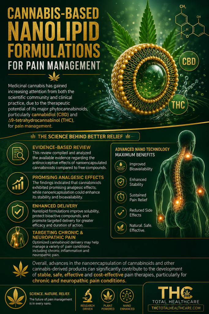
Impressive Impact of Hemp Extract on Antioxidant System in Honey Bee (Apis mellifera) Organism

“We examined the effect of hemp extract on the activity of the antioxidant system (catalase, peroxidase, glutathione, superoxide dismutase, and total antioxidant capacity) in the hemolymph of adult honey bees (Apis mellifera).

The bees were divided into three groups: (1) an experimental group fed with pure sugar syrup with cotton strips soaked with hemp extract put inside the cage; (2) an experimental group fed with a mixture of sugar syrup with hemp extract; and (3) a control group fed with a mixture of sugar and a water-glycerine solution. Hemolymph samples were collected on the 1st day of this study and then every week, until all bees in the group died.

The activities of all antioxidant enzymes were higher for the experimental groups, compared to those for the control group. The highest antioxidant activities were noted in the group supplemented with cannabis with the use of syringes. Supplementation with hemp also increased the lifespan of bees in this group compared to that of the bees consuming only sugar syrup (control: 35 days), with 49 and 52 days for groups of cannabis on strips and in syrup, respectively.

Hemp extract, thanks to its antioxidant properties, increased the activities of key antioxidant enzymes that protect the bee’s organisms against free radicals and thus delay the aging processes.”

https://pubmed.ncbi.nlm.nih.gov/35453392

“The aim of our research was to determine the effect of hemp extract on the activities of enzymes in the antioxidant system in bees’ hemolymph, along with the aging processes.”

“Hemp extract significantly increased the activity of antioxidant enzymes, extending the life of bees to 49 days (for the strip method) and 56 days (for the syringe method). In addition, we showed that a faster and stronger effect was obtained during supplementation in syrup in syringes, where the activities for the enzymes SOD, CAT, GPx, GST, and TAC were the highest.

Thanks to this, we believe that hemp extract can in the future contribute to the improvement of the natural immunity of honey bees and help them with the fight against environmental pollution and the increase of oxidative stress.”

https://www.mdpi.com/2076-3921/11/4/707

Cannabidiol (CBD) Supports the Honeybee Worker Organism by Activating the Antioxidant System

“In the experiment, we tested the effect of 30% CBD oil on the activity of the antioxidant system (superoxide dismutase, catalase, glutathione peroxidase, glutathione), the level of total antioxidant capacity, and the concentrations of ions (calcium, magnesium, and phosphorus) in honeybee workers in the hive test.

For this purpose, we prepared hives containing all stages of the development of honey bees and started the experiment by adding 200 marked, one-day old bees to each colony (intended for hemolymph collection). In the test, we created three groups (two colonies per group): (1) Experimental with CBD oil mixed with sugar syrup (CSy); (2) experimental with CBD oil on textile strips (CSt); and (3) control with pure sugar syrup only (C). Every week, we collected hemolymph from the marked bees.

In the experiment, all antioxidant enzyme activities were higher for the experimental groups CSy and CSt compared to group C. The highest concentrations/levels were obtained for the CSy group. Concentrations of calcium, magnesium, and phosphorus ions were also higher for the experimental groups compared to the C group (the highest concentration for the CSy group).

We conclude that CBD oil positively contributes to stimulating the antioxidant system of honeybees.”

https://pubmed.ncbi.nlm.nih.gov/36829838

“Our previous studies have shown that CBD oil/hemp extracts added to the diet of bees prolonged the life of insects and contributed to a positive stimulation of the immune system”

“The aim of this study was to evaluate the effect of a commercial hemp extract in the form of CBD oil on the activity of the antioxidant system”

“After a series of studies in cages and in colony conditions, we found that supplementation with CBD will potentially support the immune system of honeybees through stimulating the antioxidant system (protection against oxidative stress affecting cells and their biochemistry).

Depending on the need, the effects can be obtained regardless of the method of administration, but for the best results, we suggest using CBD in nutritional supplements (direct, faster action). In addition, research confirms that the active substance CBD may be responsible for the positive effect of the hemp extract.”

https://www.mdpi.com/2076-3921/12/2/279

CBD Supplementation Has a Positive Effect on the Activity of the Proteolytic System and Biochemical Markers of Honey Bees (Apis mellifera) in the Apiary

“We examined how CBD extract influences the activity of the immune system in the hemolymph of honey bees in the hive test. The bees were divided into 3 groups: (CSy) bees fed with CBD in sugar syrup with glycerin; (CSt) cotton strip with CBD placed in hive bees fed pure sugar syrup, (C) control bees fed sugar syrup with glycerin.

CBD extract increased the total protein concentrations, proteases and their inhibitor activities in each age (the except for acidic protease activities in the 21st and 28th day and alkaline protease inhibitor activities in the 28th day in CSt group) in comparison with group C. In the groups with the extract there was also an increase in the enzymatic marker activities: ALP, AST (decrease on day 28 for CSt), ALT; and non-enzymatic marker concentrations: glucose; triglycerides; cholesterol and creatinine. The urea acid and albumin concentrations were lower in CSy and CSt groups compared to the C group (higher concentration of albumin was displayed by control bees). Higher activities/concentrations of most of biochemical parameters were obtained in the CSy compared to the CSt and C.

CBD supplementation can positively influence workers’ immune system.”

https://pubmed.ncbi.nlm.nih.gov/36139174

“The purpose of our study was to determine how CBD extract influences resistance in the hemolymph (insect blood) of honey bees in the hive test. The bees were divided into 3 groups: (CSy) bees fed with CBD in sugar syrup; (CSt) cotton strip with CBD placed in hive, (C) control bees fed sugar syrup. To determine the state of immunity, we used the analysis of the activity of the proteolytic system and biochemical markers, such as “liver tests”, and the concentration of selected ions and key compounds for the functioning of the organism.

CBD extract increased the total protein concentration, proteases and their inhibitor activities in each age (except for acidic protease activities in the 21st and 28th day and alkaline protease inhibitor activities in the 28th day in the CSt group), increased concentrations of markers: ALP, AST, ALT; and glucose; triglycerides; cholesterol and creatinine. A decrease in concentration in experimental groups was noticed for urea acid and albumin compared to group C. Higher activities/concentrations of most of parameters were obtained in the CSy compared to the CSt and C.

The CBD supplementation can positively influence bees’ resistance.”

“CBD extract may prove to be a good supplement and can have positive effect on the immune system of honeybees by stimulating the proteolytic system and other metabolic parameters.”

https://www.mdpi.com/2076-2615/12/18/2313

Computational Characterization of Nabilone-Induced Disruption of the CB2-HER2 Receptor Complex in HER2+ Breast Cancer

“Human epidermal growth factor receptor 2-positive (HER2+) breast cancer, accounting for 15% to 20% of cases, is often resistant to treatment.

Delta-9-tetrahydrocannabinol (THC) disrupts HER2-cannabinoid receptor (2CB2) receptor complexes and inhibits HER2 activation.

This study evaluates whether Nabilone, a synthetic cannabinoid, can similarly disrupt HER2-CB2 interactions.

A CB2-HER2 complex model was generated via protein-protein docking. Three 1-µs molecular dynamics simulations (CB2-HER2, CB2-HER2-THC, CB2-HER2-Nabilone) were performed using the Schrodinger Desmond with membrane embedding and solvent. Structural stability (root mean square deviation [RMSD] and root mean square fluctuation [RMSF]), binding free energy (molecular mechanics/generalized born surface area [MM/GBSA]), and intracellular/extracellular distances between receptors were analyzed. Intermolecular interactions were assessed using the MAPIYA server.

Nabilone induced comparable structural instability to THC, with increased RMSD and RMSF. The MM/GBSA analysis showed Nabilone increased the binding free energy between CB2 and HER2, indicating stronger disruption. Intracellular and extracellular distances between CB2 and HER2 increased, especially intracellularly, with Nabilone. Intermolecular interaction analysis revealed that Nabilone decreased the number of contacts, particularly hydrophobic interactions, between CB2 and HER2.

Our in silico model predicts that Nabilone may disrupt the HER2-CB2 complex, suggesting a hypothesis that it could serve as a potential therapeutic agent. These computational findings warrant urgent experimental validation.”

https://pubmed.ncbi.nlm.nih.gov/42164472

“Cannabinoids have produced antitumor responses in preclinical models of cancer, including HER2+ BC, via binding and activating cannabinoid receptors, CB1 and CB2, both G-protein coupled receptors (GPCRs).”

“Nabilone, a synthetic analog of THC, was Food and Drug Administration (FDA)-approved in 1985 as a relief treatment for chemotherapy-related side effects, such as vomiting and nausea.”

“Our results indicate that Nabilone effectively disrupts the oncogenic CB2-HER2 complex, weakening the heterodimer interface through a mechanism of structural instability similar to THC but with superior binding affinity to CB2. While these findings rely on in silico predictions, limited by simulation timescales and simplified membrane models, they highlight a distinct opportunity for repurposing Nabilone from symptom management to active cancer therapy. We conclude that these data provide a robust theoretical framework that justifies urgent experimental validation in living systems to confirm the therapeutic potential of disrupting CB2-HER2 signaling.”

https://journals.sagepub.com/doi/10.1177/11779322261449332

Protective Effects of the Phytocannabinoid Cannabidiol on Disuse-Induced Muscle Atrophy through Modulation of Proteolysis and Mitochondrial Regulation

“Muscle atrophy induced by prolonged inactivity (disuse), including denervation-induced atrophy, is accompanied by oxidative stress, inflammation, and dysregulated protein turnover, yet no effective pharmacological therapy is currently available.

Cannabidiol (CBD), a non-psychoactive phytocannabinoid derived from Cannabis sativa, has been reported to exhibit anti-inflammatory and antioxidant properties; however, its potential involvement in disuse-related muscle atrophy has not been fully characterized.

In this study, to evaluate the potential effects of CBD on disuse-related muscle atrophy, we employed both in vivo and in vitro models. A mouse model of sciatic nerve resection-induced muscle atrophy was used for the in vivo experiments, while C2C12 myotubes were utilized for the in vitro analyses.

In the denervated mouse model, CBD attenuated the decrease in muscle mass in the tibialis anterior and gastrocnemius muscles, as well as the decline in treadmill running performance. CBD also reduced oxidative stress and suppressed the denervation-induced upregulation of Atrogin-1 and muscle RING-finger 1 (MuRF1) proteins, as well as tumor necrosis factor-α (TNF-α) mRNA.

Furthermore, CBD partially restored the decreased mitochondrial markers observed following denervation. In vitro, CBD similarly suppressed MuRF1 and Atrogin-1 protein levels and TNF-α mRNA expression in C2C12 myotubes.

These findings suggest that CBD is associated with protective effects against disuse-related muscle atrophy, accompanied by reductions in oxidative stress markers, alterations in proteolytic pathways, and changes in mitochondrial-related markers.

This study highlights a previously underexplored biological effect of a natural phytocannabinoid and supports further investigation of CBD as a potential supportive strategy for disuse-related muscle wasting.”

https://pubmed.ncbi.nlm.nih.gov/42161484

https://www.jstage.jst.go.jp/article/bpb/49/5/49_b26-00020/_article


High-molecular-weight hemp-derived polycannabidiol carbonate thermoplastic with PET-like heat resistance, strength, and processability

“Replacing polyethylene terephthalate (PET) remains a sustainability challenge because few bio-based materials match PET’s combination of low cost, high glass transition temperature (Tg), and stretch processability needed for industrial film, packaging, and bottle production.

Since PET is produced on a massive scale, multiple polymers from different renewable feedstocks will likely be necessary to significantly cut emissions linked to the global PET market. Polyethylene furanoate (PEF) is a promising alternative with similar properties, but its monomers come from food-derived sugars, connecting production to food crop supply chains and land use while requiring several chemical conversion steps that add to carbon emissions. Therefore, finding additional alternatives from non-food feedstocks is important for diversifying renewable supply options.

Here, we introduce polycannabidiol carbonate (pCBDC), a 92% bio-based thermoplastic synthesized from cannabidiol (CBD) extracted from hemp biomass, serving as a non-food, renewable, PET-like engineering plastic. pCBDC exhibits high molecular weight, high Tg, excellent stretch processability, and high strength. We also establish processing-structure-property relationships that offer guidelines for future industrial manufacturing.”

“This work demonstrates that CBD, a non-food aromatic compound extracted from hemp, can be used directly as a monomer without chemical modification to create a polymer with PET-like Tg, mechanical strength, and processability, providing a complementary pathway to PEF to reduce emissions from PET-scale materials.”

“This work presents pCBDC as a bio-based thermoplastic derived from hemp-based CBD, emphasizing its potential as a sustainable alternative to petroleum-based engineering polymers.”

“These results position pCBDC as a potential alternative to PET, polystyrene (PS), and poly(methyl methacrylate) (PMMA).”

https://www.cell.com/chem-circularity/fulltext/S3051-2948(26)00014-9

The association between prenatal exposure to cannabis with and without tobacco and early cognitive and language development in a sample of polysubstance-exposed children

Background: Prenatal cannabis exposure may have adverse effects on development which could be amplified by co-exposure with tobacco.

Objective: This study examined whether prenatal cannabis exposure was associated with disrupted language or cognitive development, and whether co-exposure to tobacco was associated with worse outcomes than to cannabis alone.

Methods: In this historical cohort study, we compared children from the Danish Family Outpatient Clinics who had prenatal exposure to cannabis (n = 106), tobacco (n = 138), cannabis and tobacco (n = 112), or no exposure to either drug (control group, n = 454) on the Bayley-III Language and Cognitive scales at 1-58 months of age (99.7% at ≤36 months). Roughly half were tested at multiple ages, yielding 1362 language assessments (49.2% tested ≥ twice) and 1549 cognitive assessments (53.6% tested ≥ twice). Most children had additional prenatal exposure to other drugs (49.8%), including the control group (63.9%). Scores were investigated in linear mixed models with factors Exposure Group, Age, and Exposure Group ∗ Age, and covariates alcohol exposure, other drug exposure, maternal medical diagnoses, and maternal education.

Results: Children with prenatal cannabis exposure scored significantly higher on the Language scale compared with controls (3.26 points; 95% CI = 0.26-6.26). There was a positive association between Language scores and age at assessment for children with cannabis and tobacco exposure (0.31 points/month; 95% CI = 0.11-0.51) but not the other groups. There were no significant effects of Exposure Group or Exposure Group × Age for the Bayley-III Cognitive scale.

Conclusion: Prenatal exposure to cannabis, alone or with tobacco, was not associated with disrupted cognitive or language development during the first three years of life in this sample of high-risk children.”

https://pubmed.ncbi.nlm.nih.gov/41990599

“Cannabis exposure was not associated with impaired cognitive or language development.”

“Using a sample of children with prenatal drug and alcohol exposure from the Danish FOCs, this study found that children with prenatal exposure to cannabis had higher scores on the Bayley-III Language scale compared with control children, and children with prenatal exposure to cannabis and tobacco had a greater increase in Bayley-III Language scores with age compared with all other groups. There were no differences on the Bayley-III Cognitive scale between children with prenatal exposure to cannabis, tobacco, both, or neither.

These results suggest that prenatal exposure to cannabis, alone or in combination with tobacco, is not associated with disrupted cognitive or language development during the first three years of life among polysubstance-exposed children. This could help to reduce the stigma experienced by women who use cannabis during pregnancy and potentially lower the barrier for seeking help in this group.”

https://www.sciencedirect.com/science/article/pii/S0378378226000745?via%3Dihub

Effect of hot and cold processed hempseed (Cannabis sativa) meal on productive performance, egg quality, egg yolk fatty acid and blood biochemical in laying hens at 37 weeks

“This study evaluated the effects of dietary inclusion of cold- and hot-processed hempseed meal (HSM) on performance, egg quality, yolk fatty acid composition, and blood biochemical parameters in laying hens.

A total of 150 Super Nick hens (37 weeks old) were allocated to three dietary treatments with five replicates of eight birds each for 16 weeks: control (0 % HSM), 15 % cold-processed HSM (60 °C), and 15 % hot-processed HSM (120 °C). Performance traits, egg production and quality indices, yolk color, fatty acid composition (gas chromatography), and serum biochemical variables were analyzed. Data were subjected to one-way ANOVA after testing assumptions, and differences among means were considered significant at P < 0.05.

Final body weight and feed intake were unaffected (P > 0.05), whereas feed conversion ratio improved in the cold-processed HSM group compared with the control (P = 0.03).

Both HSM diets increased hen-day egg production (P = 0.01) and yolk pigmentation (Roche score and b*; P < 0.01). Hot-processed HSM increased eggshell weight, ratio, and thickness (P < 0.05), while internal egg quality traits were unchanged (P > 0.05). Yolk linoleic (C18:2n6c) and α-linolenic (C18:3n3) acids increased and oleic acid (C18:1n9c) decreased in HSM-fed groups (P < 0.05). Serum triglycerides were reduced in hens fed hot-processed HSM (P = 0.04), whereas creatinine, AST, and ALT were not affected (P > 0.05).

In conclusion, dietary inclusion of 15 % hempseed meal improves feed efficiency, egg production, shell quality, and yolk fatty acid enrichment without adverse physiological effects, with processing temperature influencing the magnitude of responses.”

https://pubmed.ncbi.nlm.nih.gov/41850065

“Hemp (Cannabis sativa L.) is an annual plant that belongs to the Cannabaceae family. Hemp is used in many areas including medicine, cellulose, cosmetics, oil industry and animal feed.”

“Dietary inclusion of hempseed meal—especially in cold-processed form—can enhance feed efficiency, egg production, eggshell quality, and yolk fatty acid composition in laying hens without negatively affecting internal egg quality or blood biochemical. Additionally, hot-processed hempseed meal contributes to lower serum triglyceride levels, supporting its role in promoting healthier lipid metabolism.”

https://www.sciencedirect.com/science/article/pii/S0032579126003196?via%3Dihub

Endocannabinoid system modulation in acute, chronic, and neuropathic pain: reviewing experimental models, clinical evidence, and nanotechnology delivery

“Chronic pain is highly prevalent and inadequately managed by current therapeutic strategies, which present significant limitations such as the development of tolerance, dependence, and cognitive impairment. Therefore, searching for new pain management strategies is an ultimate goal.

The endocannabinoid system (ECS), is a broad crucial regulatory network in central nervous system’s development and in modulating various physiological and cognitive functions. It comprises endogenous cannabinoids, cannabinoid receptors, and the enzymes governing cannabinoid production and breakdown.

Recently, cannabinoids, particularly medical cannabis, have garnered renewed interest for their possibilities in treating different medical conditions, including chronic pain.

Although the risk of lethal overdose is negligible, the prevalence of non-serious adverse effects is significant and requires careful clinical consideration. Currently, there is a paucity of sufficient efficacy and long-term safety data to fully support the systematic use of medical cannabis for chronic non-malignant pain conditions.

Further research is crucial to unlock the future potential of these approaches and to delineate essential directions for exploring the ECS and its role in pain management. Advances in nanotechnology have enabled novel delivery platforms that address key limitations of cannabinoid-based therapies.

Nanocarriers, including lipid and polymeric nanoparticles, nanoemulsions, and self-emulsifying systems, can improve cannabinoid solubility, stability, bioavailability, and targeted delivery. Through controlled release and site-specific targeting, these systems hold promise for enhancing the analgesic efficacy and safety of cannabinoid therapeutics.”

https://pubmed.ncbi.nlm.nih.gov/42154330

https://link.springer.com/article/10.1007/s11011-026-01862-4

Disrupted endocannabinoid signaling contributes to systemic inflammation in acute pancreatitis

“Acute pancreatitis (AP) is an inflammatory disease that can lead to systemic complications in severe cases. The endocannabinoid system has emerged as a potential modulator of inflammation in AP.

We investigated the role of the endocannabinoid 2-arachidonoylglycerol (2-AG) and the cannabinoid receptors CB1 and CB2 during AP.

A severity-dependent decrease in circulating 2-AG was found both in patients and a murine AP model. Restoring 2-AG – by avoiding its degradation via monoacylglycerol lipase inhibitor or direct 2-AG administration – reduced local and systemic inflammation, modulated peritoneal macrophage polarization, and mitigated lung injury. Notably, endocannabinoid system effects were consistent across sexes.

Both cannabinoid receptors were involved in disease pathophysiology.

Genetic Cnr1 knockout and pharmacological CB2 blockade showed distinct and complementary roles of both receptors in regulating inflammation, immune infiltration, and pulmonary damage.

These findings highlight a protective role for 2-AG and highlight the endocannabinoid system – and cannabinoid receptors in particular – as a promising therapeutic target to modulate inflammation and reduce systemic complications in acute pancreatitis.”

https://pubmed.ncbi.nlm.nih.gov/42153289

“These findings raise the possibility that counteracting the inflammation-driven decline in endogenous 2-AG through pharmacological intervention may represent a promising therapeutic strategy for AP.”

“Our data clearly support a role for both CB1 and CB2 receptors in the pathophysiology of AP.”

“Overall, our study supports the ECS as promising therapeutic target to reduce inflammation and systemic complications in AP.”

https://pathsocjournals.onlinelibrary.wiley.com/doi/10.1002/path.70076

“The Diminished Availability of 2-AG in Aged Synaptic Terminals is Ameliorated by a Full-Spectrum Cannabis Extract with a High THC Content. This highlights the potential of high THC content extracts as therapeutic agents for restoring the decreased 2-AG levels observed in the aging brain.”

https://pubmed.ncbi.nlm.nih.gov/41880097